
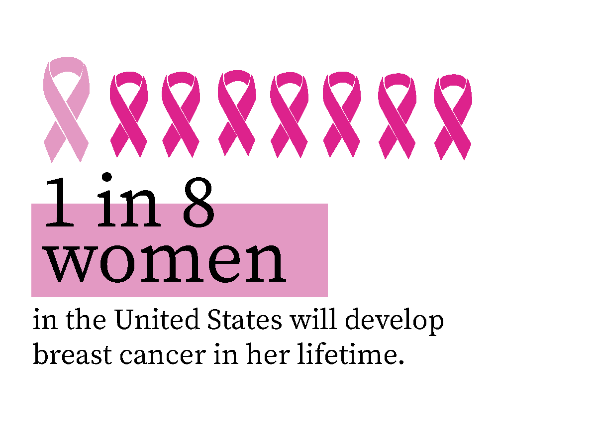
October is Breast Cancer Awareness month. One in 8 women will be diagnosed with breast cancer in her lifetime according to the National Breast Cancer Foundation. This month is a good time to encourage routine care and testing to detect early breast cancer.
Breast cancer forms in the cells of the breast. Although mostly found among women, there are some cases where it appears in men.
Symptoms of breast cancer include:
- A lump in the breast
- Change in size, shape and appearance of the breast
- Dimpling or changes to the skin over the breast
- Peeling or flaking of the skin surrounding the nipple (the areola)
- Redness or pitting of the skin over the breast
If you find a lump or other change in your breast — even if a recent mammogram was normal — don’t wait, make an appointment with your doctor.
The American Cancer Society recommends these tips about breast cancer screenings.
How often should I get a mammogram?
Women between the ages 50-74 years old are at average risk for breast cancer and can get a mammogram every two years.
It’s recommended women who are 40-49 years old should talk with their healthcare provider to discuss when they should start routine screenings and how often.
What are the different types of breast cancer screenings?
Mammogram
A mammogram is an X-ray of the breast. For many women, mammograms are the best way to find breast cancer early, when it is easier to treat and before it is big enough to feel or cause symptoms. Having regular mammograms can lower the risk of dying from breast cancer. At this time, a mammogram is the best way to find breast cancer for most women of screening age.
Breast Magnetic Resonance Imaging (MRI)
A breast MRI uses magnets and radio waves to take pictures of the breast. Breast MRI is used along with mammograms to screen women who are at high risk for getting breast cancer. Because breast MRIs may appear abnormal even when there is no cancer, they are not used for women at average risk.
Other Exams
A clinical breast exam is an examination by a doctor or nurse, who uses his or her hands to feel for lumps or other changes.
Breast Self-Awareness
Being familiar with how your breasts look and feel can help you notice symptoms such as lumps, pain, or changes in size that may be of concern. These could include changes found during a breast self-exam. You should report any changes that you notice to your doctor or health care provider.
Having a clinical breast exam or doing a breast self-exam has not been found to lower the risk of dying from breast cancer.
NEW CERIANNA PET/CT SCAN
Cerianna is a whole body new diagnostic tool that allows accurate ER characterization of metastatic lesions in a fast, non-invasive PET/CT scan. Click on the button below to learn more.