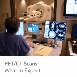
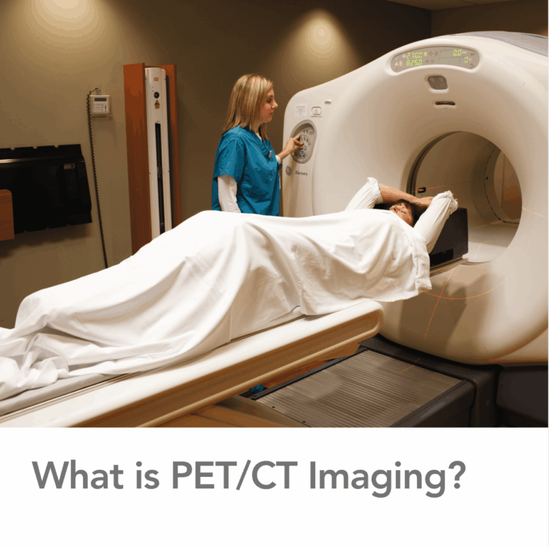
A PET/CT scan is an advanced medical imaging exam that combines two technologies—PET and CT—into a single, integrated study. This combination allows physicians to see both how the body is functioning and what it looks like structurally, all in one scan.
Understanding PET Imaging
PET imaging uses a small amount of radioactive material, called a radiotracer, to highlight areas of metabolic activity in the body. Because many diseases—especially cancer—cause changes in how cells use energy, PET scans can reveal abnormalities before structural changes occur.
Understanding CT Imaging
CT scans use X-rays to create detailed cross-sectional images of the body. These images show the size, shape, and location of organs and tissues, helping physicians pinpoint exact anatomical details.
Why Combine PET and CT?
By combining PET and CT scans:
- PET shows abnormal cellular activity
- CT shows precise anatomical location
Together, they provide a clearer, more complete picture than either scan alone.
Common Uses of PET/CT Scans
PET/CT imaging is widely used to:
- Detect and stage cancer
- Evaluate heart disease
- Assess brain disorders
- Monitor treatment response
- Identify disease recurrence
The ability to see both form and function makes PET/CT scans an essential tool in modern diagnostic imaging.
Safety and Accuracy
PET/CT scans use carefully controlled amounts of radiation and are considered safe when ordered appropriately. The benefits of accurate diagnosis and treatment planning typically outweigh the minimal risk associated with radiation exposure.
A Powerful Diagnostic Tool
PET/CT scans help physicians make informed decisions with confidence. By revealing critical details that other imaging tests may miss, PET imaging plays a vital role in early detection, accurate diagnosis, and personalized care.